
Electrochemistry of Dopamine b - hydroxylase (DBH) at a gold electrode
In cooperation with Ana Ion* and Torbjørn Ljones**
*Department of Analytical Chemistry and Instrumental Analysis, University Politehnica of Bucharest, Romania
**Department of Chemistry, Norwegian University of Science and Technology (NTNU), Trondheim, Norway
Keywords: protein, adsorption, copper-enzyme, dopamine b -hydroxylase, voltammetry
Abstract
This paper presents the results on the adsorption properties of dopamine b -hydroxylase (DBH) at a bare gold electrode. The adsorbed layer was investigated by voltammetry and electrochemical quartz crystal microbalance. Some data on the electrochemical reactivity of the copper centres in dopamine b -hydroxylase molecule are also included.
Introduction
Dopamine b - hydroxylase (DBH) (EC 1.14.17.1) is a copper enzyme which catalyzes a key hydroxylation step in the biosynthesis of noradrenaline and adrenaline, which are major hormones and neurotransmitters. Bovine adrenal enzyme DBH, which was used in this work, is a glycoproteine with the molecular weight of about 290 KD, consisting of four identical subunits. Two pairs of monomers are supposed to be held together by disulfide bridges, and each pair binds to the second one by non-covalent forces. The whole molecule contains about 30 disulfide groups, but not sulfhydryl functions. The high content of tyrosine (about 80 residues) and histidine (about 60 residues) provides a large number of sites susceptible of oxidation. By analogy with the well-characterized peptidylglycine a -hydroxylating monooxygenase (PHM) domain of peptidylglycine alpha-hydroxylating monooxygenase (E.C. 1.14.17.3), which is mechanistically similar to DBH [1], it may be inferred that each DBH molecule contains two copper atoms that take part in the catalytic reaction.
Materials and methods
DBH was extracted from bovine adrenal medulla, according to ref. [2, 3] and stored at -80 oC as an 8.7 mg/l solution in an aqueous phosphate buffer (pH 5). DBH concentration was determined by spectrophotometry at 280 nm, according to ref. [4]. After defrozing at room temperature (20 ±1 °C), a DBH sample was not used for more than 4 hours.
Apo-DBH enzyme was prepared by adding a high excess of ethylenediaminetetraacetic acid disodium salt (Complexone III, EDTA) disodium salt to the DBH solution. This mixture was left for 1 hour at room temperature, to complete the reaction.
Electrolyte solutions were prepared with fresh ultra-pure water (Millipore, 18 MW cm specific resistance). Potassium hexacyanoferrite (Merck, p.a.), ruthenium (III) hexammine chloride (Strem Chemicals, 99%) and 1,1-ferrocenedicarboxylic acid (Sigma, 96%) have been used as redox probes. Other reagents were of analytical grade A phosphate buffer (Na2HPO426 mM + NaH2PO4 20 mM + 0.1 M KNO3, pH 7), was used as supporting electrolyte, if not otherwise mentioned.
A polycrystalline gold disk electrode was prepared by embedding with Epoxy resin a gold wire (Aldrich 99.99%, 1 mm diameter) in a glass tube. Electrode surface was prepared by mechanical and electrochemical polishing [5].
Protein adsorption on the gold electrode surface was performed at room temperature (20 ±1 °C) by putting the electrode in contact with the DBH solution, for a time interval ranging between 15 and 240 minutes. The electrode was then copiously rinsed with de-ionised water, dried in N2 and transferred into the voltammetric cell.
Voltammetric experiments were performed at room temperature (20-±1 °C) by means of a PGSTAT 30 (Autolab) instrument. Dissolved oxygen was removed from the test solution by bubbling with pure nitrogen until no signal due to oxygen reduction was recorded. During the record, the nitrogen stream was directed above the test solution.
EQCM measurements have been done with a Maxtek PM-710 plating monitor.
The working electrode was a 5 MHz AT-cut quartz crystal (1-inch diameter),
coated with a Ti backed gold layer. Deposition of DBH on the piezoelectrode
surface was performed by placing the protein solution on the electrode
surface, in horizontal position. After 1 hour, the enzyme solution was
removed and the gold surface was rinsed with distilled water.
Results and Discussion
1. Behaviour of the DBH-modified gold electrode in the anodic potential region
Cyclic voltammetric scans (Fig. 1(a)) in the anodic region (potential region 0 1450 mV vs. Ag/AgCl/KCl 1M in HClO4 10 mM), corresponding to gold oxide formation (signal A), revealed an increase into the anodic charge of about 35% comparative to the charge recorded for a plain gold electrode. Also, the EQCM measurements (Fig. 1 (b)) prove that during the anodic scan the DBH surface layer is removed.
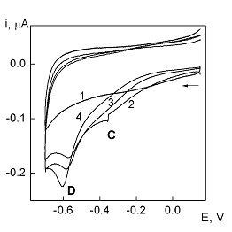
2. Cathodic reactions at DBH-modified gold electrode
Two distinct signals appear on the first scan: the shoulder C, E1/2 = - 0.30 V and the peak D, Ep = - 0.60 V (Fig. 2). An increase of the modification time (Fig. 2, curves 2 4) induces a gradual increase in the peak D current, but at the same time, the shoulder C current experiences a slight depression. Electrode processes corresponding to signals C and D are independent on each other, because a first, limited scan, in the signal C potential region does not disturb the occurrence of the process D on a second, extended scan. The anodic desorption scan performed after cathodic polarization has a similar pattern as that recorded for a fresh modified electrode. This proves the absence of desorption in the cathodic potential region.
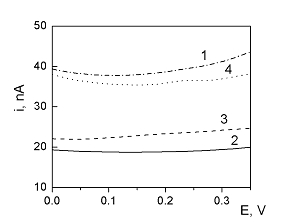
3. Capacity current measurements at DBH-modified gold electrode
DBH adsorption on the gold electrode surface induces a marked depression in the AC current (Fig. 3, curve 2). The change in capacitance is less important than that produced by a long chain alkanthiol. The capacity current does not change too much after a cathodic scan (Fig. 3, curve 3), proving that DBH is not removed from the electrode surface during the cathodic polarization.
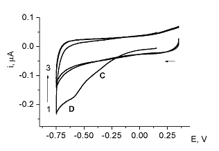
4.The electrochemical reactivity of the copper atoms in DBH
The cathodic response at DBH modified electrode (signals C and
D) was recorded also at a gold electrode modified with apo-DBH (apo-
DBH resulted after removing the copper atoms from DBH molecule by EDTA)
(Fig. 4).
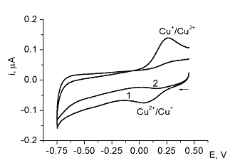
Apo-DBH is able to bind reversibly the Cu(II) ions, which are electrochemically reactive (the redox couple Cu(II)/Cu(I) gives a voltammetric response with the following characteristic peaks: Ep,c = 0.06 V ; Ep,a = 0.250 V) (Fig. 5). These atoms can be removed by contact with an EDTA solution (Fig. 5, curve 2).
Conclusions
DBH molecules adsorb on the Au electrode surface, but film formation
may involves only physical adsorption and not chemisorption through the
sulfur functions. The anodic behavior of the DBH modified gold electrode
can be assigned to the anodic oxidation of some functional groups in the
DBH molecule (histidine, imidasole). No voltammetric signal was evidenced
for the copper centers in DBH molecule. However, DBH molecules in adsorbed
state, can bind copper (II) ions from solution as was proved by the typical
voltammetric signal corresponding for Cu2+/Cu+ redox
couple (Ep,c = 60 mV; Ep,a = 250 mV).
An extended version of this work was published (ref. [6]).
References
1. S. T. Prigge, A. S. Kolhekar, B. A. Eipper, R. E. Mains, L. M. Amzel, Science, 1997, 278, 1300.
2. T. Ljones, T Skotland, T. Flatmark, Eur. J. Biochem., 1976, 61, 525.
3. T. Ljones, Meth Enzymol., 1987, 142, 596.
4. T. Ljones, T Skotland, Int. J. Peptide Protein Res, 1977, 10, 311.
5. A. Ion, V. Partali, H.-R. Sliwka, F. G. Banica, Electrochem. Commun., 2002, 4, 674 - 678.
6. Ana Ion, T. Ljones, F. G. Banica, Collect. Czech. Chem.
Commun. 69, (2004) 759.