Dirk Mohn1, Miguel Gubler1, Tobias J. Brunner1, Matthias Zehnder2, Thomas Imfeld3, Tuomas Waltimo4, and Wendelin J. Stark5. (1) Department of Chemistry and Applied Biosciences, ETH Zurich, Wolfgang-Pauli-Strasse 10, Zurich, 8093, Switzerland, (2) Department of Preventive Dentistry, Periodontology, and Cariology, University of Zürich Center of Dental Medicine, Zurich, 8093, Switzerland, (3) Departement of Preventive Dentistry, Periodontology, and Cariology, University of Zürich Center of Dental Medicine, Zurich, 8093, Switzerland, (4) Institute of Oral Microbiology and Preventive Dentistry, University of Basel, Hebelstrasse 3, Basel, 4056, Switzerland, (5) Chemistry and Applied Biosciences, ETH Zurich, Wolfgang-Pauli-Str. 10, ETH Hönggerberg, Zurich, 8093, Switzerland
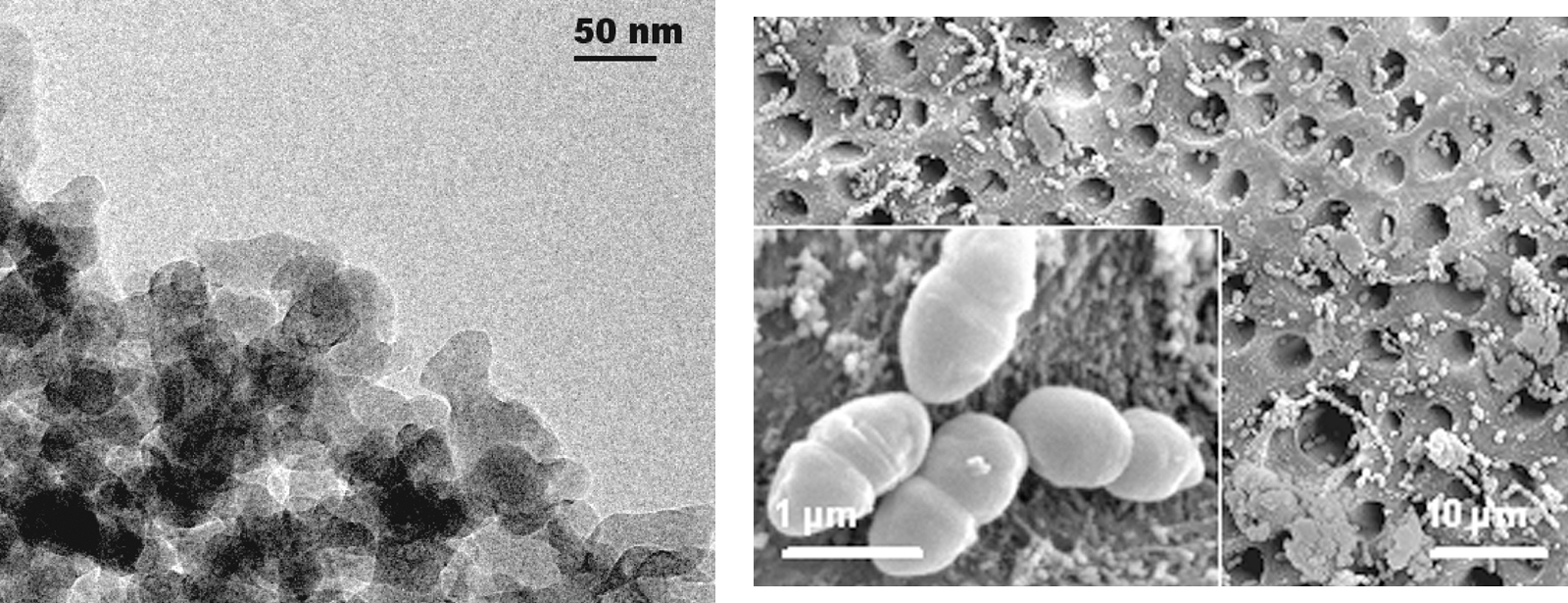
Bioactive glasses are potentially interesting materials in dentistry not only because of their ability to mineralize dentine [1] but also due to their antimicrobial effect in closed systems [2]. We investigated nanometric bioactive glasses because of their higher surface area and their faster mode of action. Furthermore, we tested the hypothesis whether nano-sized bioactive glasses also kill microbiota via mineralization or the release of ions other than sodium. Flame-spray synthesis was applied to produce nanometric glasses (Figure) with different sodium content. Calcium hydroxide was used as a control. Standardized bovine dentine disks with adherent
E. faecalis. cells were exposed to test and control suspensions.
Sodium containing glasses induced pH levels above 12, compared to less than pH 9 with sodium-free glasses. Calcium hydroxide and the sodium containing glasses killed all bacteria after 1 d. The study revealed that bioactive glasses have both, a not and a directly pH-related antibacterial effect. The former is due to ion release rather than mineralization [4].
Our study shows that nanoparticulate biomaterial can have a strong combined antimicrobial action. This opens wide applications in clinical settings.
Figure: Transmission electron microscopy image of bioactive glass nanoparticles (left). Scannning electron microscopy image of a dentine surface after treatment for 1 week with ultrafine bioactive glass showing no viable bacteria (right).
References
[1] M. Vollenweider, T.J. Brunner, S. Knecht, R.N. Grass, M. Zehnder, T. Imfeld, W.J. Stark, Acta Biomater., 2007, 3(6), 936-43.
[2] P. Stoor, E. Soderling and J.I. Salonen, Acta Odontol. Scand., 1998, 56(3), 161-5.
[3] T.J. Brunner, R.N. Grass and W.J. Stark, Chem. Commun., 2006, 13, 1384-86.
[4] M. Gubler, T.J. Brunner, M. Zehnder, T. Waltimo, B. Sener and W.J. Stark, Int. Endod. J., 2008, in press. 
